
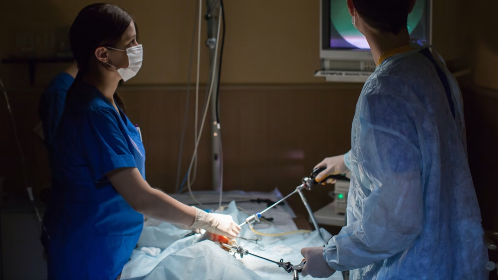
Joyvet provides veterinary endoscopy services in Bucharest for dogs and cats, including gastrointestinal endoscopy, rhinoscopy and advanced otoscopy when these procedures are medically indicated. Endoscopy allows the veterinary team to examine internal surfaces directly, collect targeted biopsy or fluid samples and, in selected cases, remove foreign material without open surgery.
Every case begins with a veterinary consultation. The initial assessment determines which procedure is appropriate, whether diagnostic imaging is needed first, how your pet should be prepared and which anesthesia or sedation protocol is safest.
This page is written for expats, international residents and visitors staying in Bucharest with their pets. When booking, please mention that you prefer to communicate in English and briefly describe your pet’s symptoms.
Veterinary endoscopy services at a glance
| Procedure | Area examined | Common reasons for referral | What may be performed |
| Gastrointestinal endoscopy | Esophagus, stomach, accessible proximal duodenum, rectum or colon | Recurrent vomiting, regurgitation, swallowing difficulty, chronic diarrhea, weight loss, gastrointestinal bleeding or a suspected swallowed object | Direct examination, targeted biopsies and selected foreign-body removal |
| Rhinoscopy | Nasal passages and nasopharynx | Persistent or one-sided nasal discharge, chronic sneezing, nosebleeds, noisy nasal breathing, facial swelling or suspected nasal foreign body | Direct examination, lavage, biopsies and selected nasal foreign-body removal |
| Advanced otoscopy or video otoscopy | External ear canal and eardrum | Recurrent otitis, ear pain, discharge, odor, head shaking, scratching, suspected grass awn, polyp or ear canal mass | Deep examination, sampling, lavage, removal of selected foreign bodies or cerumen plugs and biopsy of accessible masses |
Digestive endoscopy is primarily used for persistent gastrointestinal signs or reachable foreign bodies, rhinoscopy for persistent nasal disease, and advanced otoscopy for painful, recurrent or obstructive ear conditions.
What is veterinary endoscopy?
Veterinary endoscopy is a minimally invasive procedure that uses a flexible or rigid instrument equipped with a light source and a small video camera. The endoscope is usually introduced through a natural opening, such as the mouth, nose, rectum or ear canal, allowing the veterinarian to see areas that cannot be adequately examined from the outside.
Unlike an X-ray or ultrasound scan, endoscopy provides a real-time view of the surface of the tissue. Fine instruments can be passed through the working channel of the endoscope to collect biopsies, retrieve foreign material, perform lavage or obtain samples for laboratory testing.
Endoscopy does not replace every other diagnostic test. It shows the surfaces that the camera can reach, but it may not reveal disease located deep within an organ wall, outside the gastrointestinal tract or in inaccessible parts of the intestines and sinuses. In such cases, ultrasound, radiography, CT, MRI or surgical full-thickness biopsies may still be required.
Gastrointestinal endoscopy for dogs and cats in Bucharest
Gastrointestinal endoscopy may include:
- Esophagoscopy, for examination of the esophagus;
- Gastroscopy, for examination of the stomach;
- Upper gastrointestinal endoscopy, which may also reach the first accessible part of the duodenum;
- Colonoscopy, for examination of the rectum and colon after appropriate bowel preparation.
The procedure is performed under general anesthesia so that the patient remains still, does not feel discomfort and can be examined safely. The camera provides a magnified view of the gastrointestinal lining, including inflammation, erosions, ulcers, abnormal growths, narrowing of the lumen and foreign objects.
When may digestive endoscopy be recommended?
A veterinarian may consider endoscopy when a dog or cat has:
- recurrent or chronic vomiting;
- frequent regurgitation;
- difficulty swallowing;
- unexplained weight loss;
- persistent loss of appetite;
- chronic diarrhea;
- blood in vomit or stool;
- suspected esophagitis, gastric ulcer or gastrointestinal mass;
- a suspected object lodged in the esophagus or stomach;
- persistent digestive symptoms that remain unexplained after blood tests, radiographs or ultrasound.
Endoscopy is particularly valuable when tissue samples are needed. Even when the gastrointestinal lining looks relatively normal to the camera, microscopic examination of multiple biopsies may reveal clinically important inflammation or other disease.
Endoscopic biopsies
Small biopsy forceps can be passed through the endoscope to collect targeted samples from the esophagus, stomach, duodenum or colon. These samples are sent for histopathological examination.
Endoscopic biopsies can help differentiate between chronic inflammatory disease, certain infections, polyps and some gastrointestinal tumors. However, they mainly sample the superficial mucosal layer. When disease is suspected deeper in the intestinal wall, a full-thickness surgical biopsy may occasionally be necessary.
Endoscopic foreign-body removal for dogs and cats
Dogs commonly swallow pieces of toys, fabric, bones, socks, plastic, stones or other household objects. Cats may ingest thread, string, hair ties, rubber bands or small toys. When an object remains in the esophagus or stomach and can be reached safely, endoscopic foreign-body removal may avoid an abdominal or thoracic operation.
Once the object is visualized, the veterinarian may use forceps, snares, retrieval baskets or other instruments passed through the endoscope. The object is grasped and carefully removed through the mouth.
Not every swallowed object can be removed endoscopically. Success depends on:
- its location;
- size and shape;
- whether it is sharp or fragile;
- how long it has been present;
- whether it has already passed into the small intestine;
- whether obstruction, perforation or severe tissue damage is suspected.
Published veterinary evidence confirms that many esophageal and gastric foreign bodies can be removed endoscopically, but conversion to surgery is sometimes necessary.
Seek urgent veterinary help if your pet suddenly develops excessive drooling, gagging, repeated attempts to swallow, regurgitation, persistent vomiting, abdominal pain, refusal of food, marked lethargy or breathing difficulty after possibly swallowing an object. An esophageal foreign body should be removed promptly, while an intestinal obstruction can become life-threatening. Do not try to induce vomiting unless a veterinarian specifically instructs you to do so.
Dog and cat rhinoscopy in Bucharest
Rhinoscopy, also called veterinary nasal endoscopy, allows the veterinarian to examine the inside of a dog’s or cat’s nasal passages using a small camera. The procedure is performed under general anesthesia because complete immobility is essential for a safe and detailed examination.
Rhinoscopy may be recommended for:
- nasal discharge that persists or repeatedly returns;
- discharge affecting mainly one nostril;
- frequent or prolonged sneezing;
- recurrent reverse-sneezing episodes;
- repeated nosebleeds;
- noisy nasal breathing or nasal obstruction;
- facial swelling or asymmetry around the nose;
- suspicion of a grass awn, seed or other nasal foreign body;
- suspicion of chronic rhinitis, fungal disease, a polyp or a nasal mass.
Persistent unilateral discharge is especially important because it may indicate a localized foreign body, fungal lesion or mass rather than a generalized respiratory infection.
What can rhinoscopy find?
Rhinoscopy may reveal:
- inflamed or abnormal nasal tissue;
- accumulated mucus, pus or blood;
- fungal plaques;
- grass awns, seeds and other foreign material;
- nasal polyps;
- abnormal masses;
- structural narrowing or scar tissue;
- areas from which a targeted biopsy should be collected.
If a reachable foreign body is found, the veterinarian may be able to grasp and remove it during the same procedure. Suspicious tissue can be sampled for histopathology, while secretions or abnormal material may be collected for additional laboratory testing.
Rhinoscopy and CT are complementary
Rhinoscopy shows the visible surface of the nasal cavity and permits targeted biopsy or foreign-body retrieval. CT provides a cross-sectional view of the skull, nasal bones, sinuses and surrounding structures.
When a deep mass, extensive fungal disease or sinus involvement is suspected, CT may be recommended before rhinoscopy to define the location and extent of the lesion. Rhinoscopy can then be used to inspect the accessible area and obtain samples. Research comparing these methods supports using CT and rhinoscopy as complementary rather than interchangeable investigations.
Veterinary otoscopy and video otoscopy in Bucharest
A routine otoscopic examination uses a handheld illuminated instrument to inspect the entrance and accessible part of the ear canal. Many cooperative pets can undergo this examination while awake during a normal veterinary consultation.
Advanced or video-assisted otoscopy is more appropriate when the canal must be examined deeply, the problem is painful or recurrent, the canal contains large amounts of discharge, or lavage, sampling, foreign-body removal or biopsy is planned.
When should your pet’s ears be examined?
Otoscopy is recommended when a dog or cat has:
- repeated head shaking;
- scratching or rubbing of the ears;
- ear pain;
- redness or swelling;
- excessive wax or abnormal discharge;
- an unpleasant odor from the ear;
- recurrent infection in the same ear;
- apparent hearing loss;
- head tilt, imbalance or other vestibular signs;
- suspicion of an ear mite, grass awn, polyp or tumor.
The veterinarian examines the external ear canal and, when visible, the tympanic membrane or eardrum. The appearance and integrity of the eardrum influence which cleaning products and medications can be used safely.
What may be done during advanced otoscopy?
Depending on the case, advanced otoscopy may allow:
- collection of material for cytology;
- bacterial culture and susceptibility testing;
- controlled ear lavage;
- removal of cerumen plugs;
- removal of grass awns or other foreign bodies;
- assessment of a perforated or abnormal eardrum;
- visualization and biopsy of polyps or ear canal masses.
Video otoscopy improves visualization and can assist with foreign-body retrieval, biopsy and procedures involving the eardrum.
Simple otoscopy may not require sedation. Sedation or general anesthesia is more likely to be recommended when the ear is severely painful, the patient cannot remain still or a deep lavage, foreign-body extraction or biopsy is needed.
Advanced otoscopy helps diagnose and treat the immediate ear problem, but recurrent otitis still requires investigation of the underlying cause. In dogs, this may include allergy or another dermatological disorder. Technology cannot compensate for an incomplete medical work-up of the primary disease.
Endoscopy, ultrasound, X-ray or CT: which test does your pet need?
These investigations answer different questions and are often used together.
| Investigation | Main strength | Important limitation |
| X-ray | Can identify some radiopaque objects, major bony changes and indirect signs of obstruction | Does not show the mucosal surface and can miss radiolucent foreign material |
| Ultrasound | Examines abdominal organs, intestinal wall thickness, lymph nodes, fluid and surrounding tissues | Does not provide a direct view of the internal mucosal surface and cannot collect endoscopic biopsies |
| CT | Defines the location and extent of deep nasal, skull or other cross-sectional lesions | Does not provide histopathological confirmation and cannot directly remove most lesions |
| Endoscopy | Directly visualizes accessible internal surfaces and allows targeted sampling or selected treatment | Cannot reach every part of the intestine or sinus and may sample only the superficial tissue layer |
Radiography and ultrasound are often appropriate first-line tests. Endoscopy becomes especially useful when the abnormality is suspected at the mucosal surface, targeted biopsy is required or a reachable foreign body may be removed. A normal ultrasound does not automatically exclude small ulcers or subtle mucosal disease.
For the other investigations discussed here, see our guide to pet X-rays, ultrasound and blood tests.
What happens before, during and after veterinary endoscopy?
1. Initial consultation
The veterinarian reviews your pet’s symptoms, previous diagnoses, current medication and relevant blood tests or imaging. Depending on age and health status, pre-anesthetic blood work, chest imaging, cardiac assessment or additional investigations may be recommended.
For international patients, bring or send any available:
- medical reports;
- laboratory results;
- X-rays, ultrasound or CT images;
- medication list and doses;
- histopathology or cytology reports;
- videos showing coughing, sneezing, regurgitation or abnormal breathing.
2. Preparation
Preparation depends on the procedure.
For upper gastrointestinal endoscopy, fasting is normally required so that the stomach is empty. Colonoscopy requires more extensive bowel preparation, which may include a special diet, laxatives or enemas.
Rhinoscopy is also generally performed after appropriate fasting because general anesthesia is required.
For an ear examination, do not clean deeply inside the ear before the appointment unless instructed otherwise. Cleaning can remove diagnostically useful material or push a foreign body further into the canal.
Always follow the individual instructions provided by the clinic. Fasting requirements may be modified for very young pets, diabetic patients or animals with other medical conditions.
3. Anesthesia or sedation
Gastrointestinal endoscopy and rhinoscopy are generally performed under general anesthesia. This protects the patient, allows precise instrument control and prevents pain or sudden movement.
The need for sedation or anesthesia during otoscopy depends on the degree of pain, the patient’s temperament and the depth of the planned examination or intervention.
4. Direct examination, sampling and treatment
The veterinarian systematically examines the target area. Images and findings are recorded, and samples are collected from abnormal sites when indicated.
When a suitable foreign body is found, removal may be attempted during the same anesthetic episode. However, the veterinarian may stop the attempt and recommend surgery if continued manipulation could damage the tissue.
5. Recovery and results
Many stable patients can return home on the same day after recovering sufficiently from anesthesia. Hospitalization may still be recommended if the pet is medically unstable, the procedure was complicated or continued monitoring is required.
The veterinarian can usually discuss the visual findings shortly after the procedure. Biopsy results are not immediate because the samples must be processed and examined by a laboratory. Turnaround time depends on the laboratory and the type of analysis requested.
Is veterinary endoscopy safe?
Endoscopy is minimally invasive, but no anesthetic or medical procedure is completely risk-free. The individual risk depends on the patient’s age, breed, general health, existing heart or respiratory disease and the exact procedure being performed.
Possible short-term effects include:
- temporary sleepiness after anesthesia;
- mild throat irritation after upper gastrointestinal endoscopy;
- temporary nasal irritation or blood-streaked discharge after rhinoscopy;
- short-lived ear discomfort after deep otoscopy or lavage.
A small amount of nasal bleeding may occur after rhinoscopy, particularly when biopsies have been collected. Heavy or persistent bleeding, marked breathing difficulty, repeated vomiting or severe pain should be reported to the veterinary team immediately.
The veterinary team assesses the expected benefit against the anesthetic and procedural risks before recommending endoscopy.
How much does dog or cat endoscopy cost in Bucharest?
The final veterinary endoscopy cost in Bucharest depends on:
- the type of procedure;
- the duration and complexity of the case;
- the anesthesia or sedation protocol;
- pre-anesthetic blood tests;
- radiographs, ultrasound or CT;
- biopsy collection;
- histopathology, cytology or microbiology;
- lavage or foreign-body removal;
- medication and postoperative monitoring.
Joyvet publishes separate prices for gastrointestinal endoscopy, endoscopic foreign-body removal, anterior and retrograde rhinoscopy, otoscopy, sample collection and lavage. Published procedure prices do not include anesthesia unless specifically stated. For an accurate estimate, the patient must first be assessed and the complete diagnostic plan established.
Veterinary endoscopy for expats and international pet owners in Bucharest
When arranging an appointment from abroad or while temporarily staying in Romania, provide as much medical information as possible before the visit. Previous records reduce unnecessary repetition and help the veterinarian decide whether your pet needs endoscopy immediately or whether another investigation should come first.
When contacting Joyvet, include:
- your pet’s species, breed, age and weight;
- the main symptoms;
- when the symptoms began;
- whether they are constant or intermittent;
- whether a foreign object may have been swallowed or inhaled;
- previous investigations and treatments;
- known illnesses or previous anesthetic reactions;
- your preferred language for communication.
Please mention that you would prefer to communicate in English when booking. Learn more about our English-speaking veterinary clinic in Bucharest.
Joyvet Veterinary Clinic
Call: +40731803803
WhatsApp: +40731803803
Address: 68 Dumitru Papazoglu Street, Sector 3, Bucharest
Email: joyvetbucuresti@gmail.com
Frequently asked questions
Where can I book veterinary endoscopy in Bucharest?
Joyvet offers gastrointestinal endoscopy, rhinoscopy and advanced otoscopy for dogs and cats when medically indicated. The first step is a consultation to determine the correct procedure, required preparation and anesthetic plan.
Does my dog or cat need general anesthesia for endoscopy?
Gastrointestinal endoscopy and rhinoscopy generally require general anesthesia. Routine otoscopy may be possible while the animal is awake, but advanced otoscopy may require sedation or anesthesia when the ear is painful or an intervention is planned.
Can endoscopy remove a swallowed object without surgery?
Sometimes. Many objects located in the esophagus or stomach can be grasped and removed endoscopically. Surgery may still be required when the object is inaccessible, very large, embedded, associated with perforation or already located further along the intestines.
What symptoms may indicate that my dog needs rhinoscopy?
Persistent or unilateral nasal discharge, chronic sneezing, recurrent nosebleeds, noisy nasal breathing, facial swelling and sudden sneezing after walking through grass or fields may justify a rhinoscopy assessment.
Can rhinoscopy remove a grass seed from a dog’s nose?
A reachable grass awn or seed can sometimes be identified and removed through the rhinoscope. The possibility of removal depends on its location, surrounding inflammation and how long it has been present.
Does every otoscopy require sedation?
No. A cooperative animal with a mildly affected ear may be examined without sedation. Sedation or anesthesia becomes more likely when the ear is very painful, the canal is obstructed or the veterinarian needs to perform lavage, biopsy or foreign-body removal.
Should I clean my pet’s ear before the appointment?
Do not perform deep cleaning unless instructed by the veterinarian. Cleaning may remove useful diagnostic material, alter cytology results or push a foreign body further into the canal.
Can endoscopy still be useful after a normal ultrasound or X-ray?
Yes. Some diseases mainly affect the mucosal surface and may not be visible on routine imaging. Endoscopy provides direct visualization and can obtain biopsies, although it also has limitations and cannot exclude every deep lesion.
Can my pet go home on the same day?
Many patients go home after they have recovered from anesthesia and are clinically stable. The final decision depends on the procedure, findings, anesthetic recovery and the pet’s general condition.
How quickly will I receive the biopsy result?
The veterinarian can discuss the visible findings shortly after the procedure. Histopathology and other laboratory tests require additional processing time, so the definitive report normally follows later.
Book a veterinary endoscopy assessment in Bucharest
Persistent vomiting, regurgitation, chronic nasal discharge, repeated nosebleeds, recurrent ear infections and suspected foreign bodies should not be treated indefinitely without identifying the underlying cause.
Contact Joyvet to arrange an assessment for gastrointestinal endoscopy, dog or cat rhinoscopy, advanced otoscopy, targeted biopsy or selected endoscopic foreign-body removal.
For suspected foreign-body ingestion, marked breathing difficulty, severe nasal bleeding, excessive drooling, repeated gagging or an inability to swallow, call the clinic before travelling so the case can be triaged appropriately.